Epithelioid mesothelioma pathology outlines are crucial for understanding this aggressive cancer that affects the mesothelial cells lining various organs. As the most common subtype of mesothelioma, epithelioid cases demand precise diagnosis and tailored treatment approaches. This guide delves into the intricate world of epithelioid mesothelioma pathology, exploring its unique cellular characteristics, diagnostic challenges, and implications for patient care. Whether you’re a healthcare professional or someone seeking to understand this complex condition, we’ll unravel the pathological features that make epithelioid mesothelioma distinct and discuss how these insights shape treatment strategies and patient outcomes.
Key Takeaways:
- Epithelioid mesothelioma is the most common subtype of this rare cancer
- Accurate pathological diagnosis is crucial for effective treatment planning
- Distinctive cellular patterns and immunohistochemical markers aid in identification
- Advanced diagnostic techniques, including molecular pathology, are improving accuracy
- Pathological findings significantly impact prognosis and treatment options
Understanding Epithelioid Mesothelioma
Definition and Classification
Epithelioid mesothelioma is a subtype of malignant mesothelioma, accounting for approximately 50-70% of all cases. This cancer originates in the mesothelial cells lining the pleura, peritoneum, or pericardium. Unlike its sarcomatoid and biphasic counterparts, epithelioid mesothelioma is characterized by cells that closely resemble normal mesothelial cells, making it crucial for pathologists to distinguish between benign and malignant conditions.
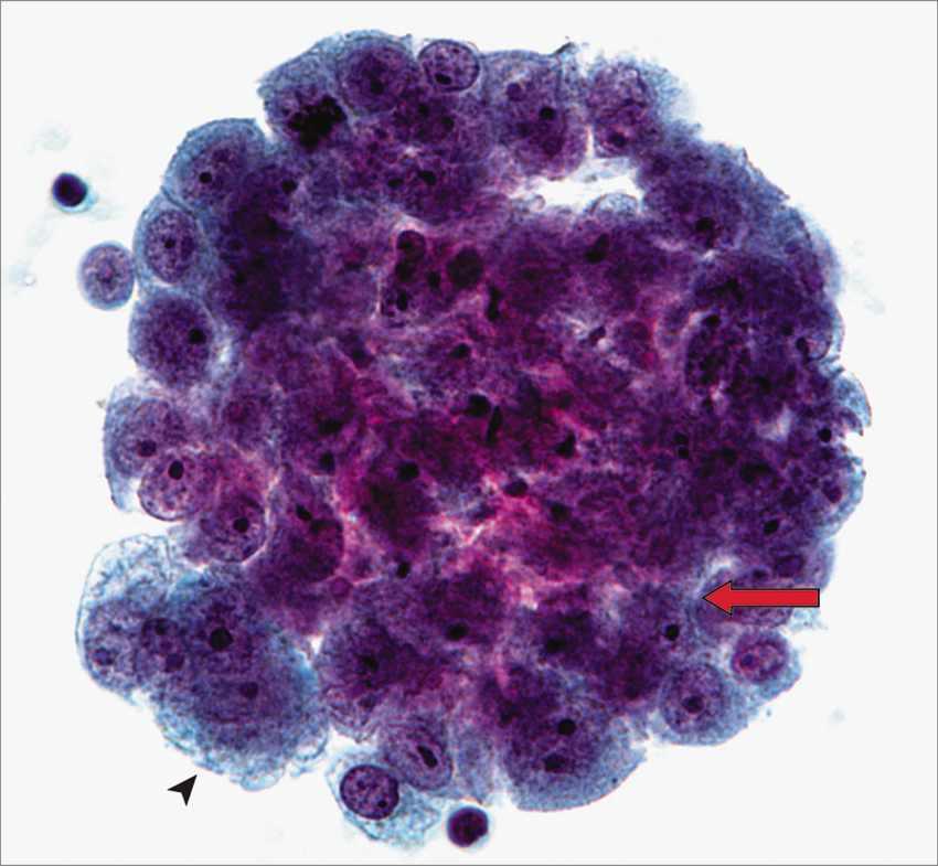
Cellular Characteristics
Epithelioid mesothelioma cells typically appear round or cuboidal with abundant cytoplasm and centrally located nuclei. These cells often form tubular, papillary, or sheet-like patterns, which are visible under microscopic examination. Compared to normal mesothelial cells, epithelioid mesothelioma cells exhibit increased nuclear-to-cytoplasmic ratios and more prominent nucleoli.
Pathological Features of Epithelioid Mesothelioma
Gross Appearance
Macroscopically, epithelioid mesothelioma presents as multiple small nodules or a diffuse thickening of the affected mesothelial surface. In advanced stages, it can form a rind-like encasement of organs. The pleura is the most common site, accounting for about 75% of cases, followed by the peritoneum (10-20%) and rarely the pericardium or tunica vaginalis.
Microscopic Findings
Under the microscope, epithelioid mesothelioma displays various growth patterns, including:
- Tubulopapillary (most common, seen in 40% of cases)
- Solid (seen in 20% of cases)
- Trabecular
- Micropapillary
These patterns can coexist within the same tumor, adding complexity to the diagnosis.
Immunohistochemistry
Immunohistochemical staining plays a crucial role in diagnosis. Key positive markers for epithelioid mesothelioma include:
- Calretinin (positive in 95% of cases)
- WT1 (positive in 90% of cases)
- CK5/6 (positive in 75-100% of cases)
- D2-40 (podoplanin) (positive in 90-100% of cases)
Negative markers like CEA, TTF-1, and Napsin-A help differentiate from adenocarcinomas.
Importance of Accurate Pathological Diagnosis
Impact on Treatment Planning
Precise pathological diagnosis is essential for tailoring treatment strategies. Epithelioid mesothelioma generally responds better to treatment compared to other subtypes. For instance, patients with epithelioid type may be candidates for aggressive surgical approaches like extrapleural pneumonectomy, which has shown a median survival of 19 months in selected cases.
Prognostic Implications
Epithelioid mesothelioma carries a better prognosis compared to other subtypes. Studies have shown a median survival of 12-27 months for epithelioid type, compared to 4-18 months for sarcomatoid and 8-21 months for biphasic types. This information is crucial for patient counseling and treatment decision-making.
Challenges in Epithelioid Mesothelioma Diagnosis
Differential Diagnosis
Distinguishing epithelioid mesothelioma from other conditions can be challenging. Key differentials include:
- Reactive mesothelial hyperplasia
- Metastatic adenocarcinoma
- Serous carcinoma (in peritoneal cases)
Advanced techniques like BAP1 immunohistochemistry and FISH for CDKN2A deletion can aid in difficult cases, with studies showing 70-80% sensitivity for malignant mesothelioma.
Sampling and Biopsy Considerations
Adequate tissue sampling is crucial for accurate diagnosis. Video-assisted thoracoscopic surgery (VATS) biopsy is preferred, offering a diagnostic yield of over 90% compared to 60-70% for CT-guided needle biopsy. Multiple biopsies from different areas are recommended to account for tumor heterogeneity.
Advanced Diagnostic Techniques
Molecular Pathology
Recent advances in molecular pathology have identified key genetic alterations in epithelioid mesothelioma:
- BAP1 mutations (present in 40-60% of cases)
- NF2 mutations (present in 35-40% of cases)
- CDKN2A deletions (present in 45-50% of cases)
These findings are not only diagnostic but also offer potential therapeutic targets.

Conclusion
Epithelioid mesothelioma pathology outlines serve as a crucial roadmap in the complex landscape of this rare but aggressive cancer. As our understanding deepens, so does our ability to tackle this formidable foe. The intricate dance of cellular patterns, immunohistochemical markers, and molecular signatures not only aids in precise diagnosis but also paves the way for personalized treatment strategies. Looking ahead, the integration of artificial intelligence in pathology analysis and the emergence of liquid biopsies promise to revolutionize how we approach epithelioid mesothelioma. These advancements may soon allow for earlier detection and more targeted therapies, potentially turning the tide in the battle against this challenging disease. As we continue to unravel the mysteries of epithelioid mesothelioma, the collaboration between pathologists, oncologists, and researchers becomes ever more critical, offering hope for improved outcomes and, ultimately, a brighter future for those affected by this rare cancer.