Malignant pleural mesothelioma radiology plays a crucial role in diagnosing and managing this aggressive cancer. As the primary tumor of the pleura, MPM poses unique challenges for healthcare professionals. With its increasing incidence linked to asbestos exposure, understanding the intricacies of radiological assessment is more important than ever. This comprehensive guide dives into the world of MPM imaging, exploring various modalities, their strengths, and limitations. We’ll unravel the complexities of diagnosis, from initial chest X-rays to advanced PET-CT scans, and discuss how these tools shape treatment decisions and patient outcomes.
Key Takeaways:
- MPM diagnosis relies on a multi-modality imaging approach
- CT scans are pivotal for identifying malignant processes
- MRI offers superior contrast resolution for local disease evaluation
- PET-CT provides crucial metabolic and anatomical insights
- Ultrasound is highly effective for monitoring pleural effusions
Understanding Malignant Pleural Mesothelioma
Malignant pleural mesothelioma (MPM) is a rare but aggressive cancer that originates in the pleura, the thin membrane lining the lungs and chest cavity. Predominantly caused by asbestos exposure, MPM has a long latency period of 30-40 years. In the United States, the incidence is approximately 15 cases per million annually for men and 3 for women, with a higher prevalence in those aged 60 to 80.
Epidemiology and Risk Factors
Asbestos exposure remains the primary risk factor, accounting for over 80% of MPM cases. Occupational exposure in industries such as construction, shipbuilding, and mining significantly increases the risk. Other factors include genetic predisposition and exposure to certain minerals like erionite.
The Role of Imaging in MPM Diagnosis
Radiological assessment is crucial for MPM diagnosis, staging, and treatment planning. A multi-modality approach utilizing various imaging techniques provides a comprehensive evaluation of the disease.
Key Imaging Modalities for MPM
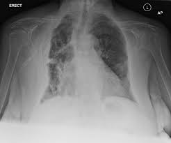
Chest Radiography (CXR)
As the first-line examination, CXR can detect unilateral pleural effusion in 30%-80% of cases and pleural-based masses in less than 25%. However, its sensitivity and specificity are limited, necessitating further imaging.
Computed Tomography (CT)
CT scans are pivotal in MPM evaluation, offering enhanced sensitivity for malignant processes. Key features include:
- Nodular or lobular pleural thickening (90%-92% of patients)
- Circumferential thickening in advanced cases
- Sensitivity of 68% for distinguishing malignant from benign conditions
Magnetic Resonance Imaging (MRI)
MRI provides excellent contrast resolution, aiding in differentiating MPM from adjacent structures. On T1-weighted scans, MPM appears iso/hypointense, while T2-weighted images show hyperintensity. MRI excels in assessing local disease extent and predicting surgical resectability.
Positron Emission Tomography-Computed Tomography (PET-CT)
PET-CT delivers both metabolic and anatomical insights, crucial for accurate staging and treatment planning. A SUV cutoff of 2.0-2.2 demonstrates sensitivities of up to 100% in distinguishing malignancies from benign pleural diseases.
Ultrasonography (US)
US is highly effective in monitoring pleural effusions and guiding interventional procedures. It boasts a specificity of 100% in differentiating malignant from benign effusions, surpassing CT in assessing complex fluid patterns.
Challenges in MPM Radiological Diagnosis
Despite advancements, MPM diagnosis faces several challenges:
- Differentiating MPM from other pleural diseases
- Limitations of individual imaging modalities
- Variability in disease presentation
Overcoming Diagnostic Challenges: Best Practices
To enhance diagnostic accuracy, consider the following strategies:
- Integrate multiple imaging techniques for a comprehensive assessment
- Implement standardized reporting protocols to ensure consistency
- Correlate radiological findings with clinical data
- Utilize image-guided biopsies for definitive diagnosis
Advanced Imaging Techniques and Future Directions
Emerging technologies are shaping the future of MPM imaging:
- Artificial intelligence and radiomics for improved detection and characterization
- Novel PET tracers for enhanced metabolic imaging
- Dual-energy CT for better tissue characterization
Impact of Radiological Assessment on Treatment
Radiological findings significantly influence MPM management:
- Guide treatment selection between surgery, chemotherapy, and radiation
- Monitor treatment response with serial imaging
- Facilitate follow-up and surveillance to detect recurrence early
In conclusion, the integration of advanced imaging techniques in malignant pleural mesothelioma radiology has revolutionized diagnosis and management. By leveraging the strengths of each modality and addressing challenges through best practices, clinicians can provide more accurate diagnoses and tailored treatment plans, ultimately improving patient outcomes in the face of this aggressive malignancy.

Paving the Way for Better MPM Management Through Advanced Radiology
As we’ve explored the intricate world of malignant pleural mesothelioma radiology, it’s clear that imaging techniques are the cornerstone of effective diagnosis and treatment. The multi-modality approach, combining CT, MRI, PET-CT, and ultrasound, offers a comprehensive view of this complex disease. Yet, the journey doesn’t end here. The future holds exciting possibilities with AI and radiomics potentially revolutionizing early detection and personalized treatment plans. As we continue to refine our understanding of MPM’s radiological signatures, we’re not just improving diagnostics – we’re opening doors to more targeted therapies and better patient outcomes. The evolving landscape of MPM radiology serves as a beacon of hope, promising more accurate staging, tailored treatments, and ultimately, improved quality of life for those affected by this challenging malignancy. In this ever-advancing field, staying informed and embracing new technologies will be key to conquering the complexities of MPM.