Cystic peritoneal mesothelioma radiology isn’t just a mouthful—it’s a crucial piece of the diagnostic puzzle for this rare abdominal condition. Whether you’re a budding radiologist or a curious patient, this guide will demystify the imaging techniques used to spot this elusive disease. We’ll dive into the world of ultrasounds, CT scans, and MRIs, unraveling the telltale signs that set CPM apart from its look-alikes. By the end, you’ll have a clearer picture (pun intended!) of how radiologists navigate the twists and turns of CPM diagnosis. So, grab your virtual lab coat, and let’s embark on this radiological adventure together!
Essential Diagnostic Arsenal for CPM
When it comes to unmasking cystic peritoneal mesothelioma, radiologists aren’t just winging it. They’ve got a powerful toolkit at their disposal. This isn’t your average game of hide-and-seek; it’s a high-stakes detective work where every pixel counts. From cutting-edge imaging machines to specialized software, each tool plays a crucial role in piecing together the CPM puzzle. Let’s peek behind the curtain and explore the gear that makes this diagnostic magic happen. Whether you’re a medical pro looking to brush up or a curious patient, understanding these tools will give you a whole new appreciation for the art and science of CPM radiology.
Imaging Modalities: The Big Three
- Ultrasound Machine: The first line of defense, offering real-time, radiation-free imaging.
- CT Scanner: Provides detailed cross-sectional views, crucial for spotting those sneaky cysts.
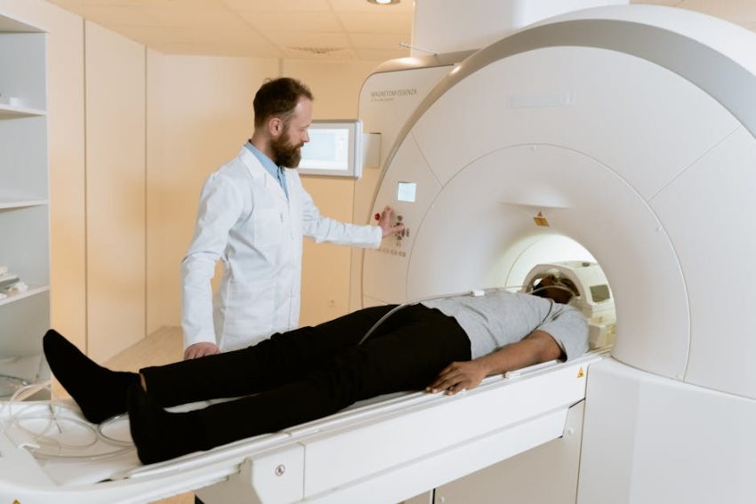
- MRI System: The heavyweight champ, delivering unparalleled soft tissue contrast.
Software and Analysis Tools
- PACS (Picture Archiving and Communication System): The digital backbone for storing and sharing images.
- 3D Reconstruction Software: Turns flat images into immersive, manipulable 3D models.
- AI-assisted Detection Programs: Cutting-edge tech that helps flag potential CPM markers.
Specialized Equipment
- Contrast Agents: These bad boys light up the show, making cysts pop on images.
- High-resolution Monitors: Because every detail matters when you’re hunting for CPM.
- Biopsy Tools: For when images alone just won’t cut it, and we need to get up close and personal.
Navigating the CPM Imaging Maze: A Step-by-Step Guide
Alright, fellow imaging enthusiasts, it’s time to roll up our sleeves and dive into the nitty-gritty of CPM detection. We’re about to embark on a radiological journey that’ll make Sherlock Holmes jealous. From initial patient prep to the final “Aha!” moment, we’ll walk you through each twist and turn of the CPM imaging process. Buckle up, because this isn’t your grandma’s X-ray – we’re talking cutting-edge tech and eagle-eyed analysis. Whether you’re a radiologist-in-training or just curious about what goes on behind those closed imaging suite doors, this guide will illuminate the path to CPM discovery.
1. Setting the Stage: Patient Prep and Initial Assessment
- Gather detailed patient history, focusing on abdominal symptoms and risk factors
- Perform physical examination, noting any palpable masses or tenderness
- Explain the imaging process to the patient, addressing any concerns
Pro Tip: A thorough patient history can be a game-changer. Don’t skimp on the details!
2. Ultrasound: The First Line of Defense
- Position patient for optimal abdominal visualization
- Conduct a systematic scan, paying extra attention to the peritoneal cavity
- Look for the telltale “spider in a web” appearance characteristic of CPM
Watch out for: Misinterpreting normal bowel loops as cystic structures. When in doubt, get a second opinion!
3. CT Scan: Peeling Back the Layers
- Administer oral and IV contrast as appropriate
- Perform multi-phase scanning to capture both arterial and venous phases
- Analyze images for low-density, thin-walled cystic masses
Key Point: Don’t forget to check for calcifications – they’re rare in CPM but can be a crucial differentiator.
4. MRI: The Detail Devil
- Select appropriate sequences (T1, T2, DWI) for comprehensive evaluation
- Look for characteristic high T2 signal intensity of cystic components
- Assess for any solid components or septations within the cysts
Pro Tip: Fat-suppressed sequences can be a game-changer in distinguishing CPM from other fatty lesions.
5. Putting It All Together: Image Interpretation
- Compare findings across all imaging modalities
- Consider differential diagnoses such as lymphangiomas or pseudomyxoma peritonei
- Correlate radiological findings with clinical presentation
Remember: CPM is a rare beast. If something looks off, don’t hesitate to consult with colleagues or seek a second opinion.

Conclusion: Unmasking the Cystic Culprit
As we wrap up our deep dive into cystic peritoneal mesothelioma radiology, it’s clear that this rare condition demands a keen eye and a multifaceted approach. From the initial ultrasound whispers to the MRI’s eloquent revelations, each imaging modality plays a crucial role in piecing together the CPM puzzle. But beyond the technical aspects, what truly sets apart exceptional CPM diagnosis is the radiologist’s ability to synthesize these findings with clinical context. It’s not just about spotting cysts; it’s about understanding their story within the patient’s broader health narrative. As imaging technology evolves, so too must our interpretative skills. The future of CPM radiology lies not just in sharper images, but in sharper minds capable of navigating the complex interplay between pixels and patient. So, whether you’re a seasoned pro or a curious learner, keep pushing the boundaries of your diagnostic acumen. The next breakthrough in CPM detection could be just one scan away.
Additional Tips and FAQs
- Tip: Always compare current images with prior studies when available. Subtle changes over time can be key in CPM diagnosis.
- FAQ: Can CPM be definitively diagnosed through imaging alone?
Answer: While imaging plays a crucial role, definitive diagnosis typically requires histopathological confirmation through biopsy. - Tip: Don’t underestimate the power of interdisciplinary collaboration. Regular tumor board discussions can significantly enhance diagnostic accuracy.
- FAQ: How often should follow-up imaging be performed in CPM patients?
Answer: Follow-up schedules vary, but typically, imaging is recommended every 3-6 months initially, then annually if stable. Always consult with the treating physician for personalized recommendations. - Tip: Stay updated on emerging AI-assisted detection tools. While not replacing human expertise, they can serve as valuable second readers in complex cases.
#cystic peritoneal mesothelioma radiology #CPM imaging #diagnostic techniques #abdominal cysts #mesothelioma detection #radiological findings #medical imaging